The Case of the Mystery Bacteria: Part II
When we left, two plates were incubating after two types of inoculation, lawn culture and isolation streak. 20 hours and 30 minutes later.......

WE HAVE GROWTH!!!
Outside of the many types of fermentation I love, this is the first time I have intentionally cultivated a bacterial colony. A clear difference can be seen between the two different inoculation techniques.
Bacteria grow in colonies that can be seen without the aid of a microscope even though the individual bacteria in the colony are microscopic. All the bacteria in the colony above are clones of the bacteria originally inoculated on the plate. Each species of bacteria produces its own unique bacterial colony with its own characteristic appearance. The description of this appearance is referred to as colony morphology. Below are some of the common ways in which colony morphology is described:

The colony morphology for the unknown bacteria:
- Form: Circular
- Elevation: Convex
- Margin: Entire
- Pigment: Yellow-ish
- Optical Characteristics: Opaque
- Consistency:
- Viscid (sticky when poked with an object)
After recording a macroscopic view of the bacteria, I prepared a slide to examine the bacteria on a microscopic scale using a..............microscope. Slides are created using a technique called smear preparation. The process is simple, but requires care as if the smear is too thin or thick it may contain too few or too many bacterial cells to stain properly (what's staining, we will get to that soon). Smear technique involves mixing a small amount of bacteria with a small droplet of water and allowing it to dry completely. Once dried heat is applied in a process called heat fixation, in which the cells are killed and adhere to the slide.
You might be thinking, why do I not just speed up the process and use heat to dry and fix the slide at the same time. Unfortunate, application of heat during the drying process will cause distortion and possible rupture of the cells to be examined due to boiling. So one must just be patient.
The circle you see in the picture below is on the opposite side of the slide. It is there as a "target" should the smear be difficult to find in the field of view of the microscope.

Now that staining I mentioned earlier. Not only does staining enhance the visibility of bacteria under the microscope, the particular stain process I performed, Gram staining, provides additional information about the identity of the bacteria. You might have keenly noticed that the "G" in Gram is capitalized. That is because the method was named after its inventor Hans Christian Gram. Gram staining helps distinguish the composition of the cell wall of a bacteria. I will go into details about the biology of this in a separate post. At the moment it is important to know that the most common human pathogenic bacteria have either a Gram-positive cell wall with multiple peptidoglycan layers, or a Gram-negative cell wall with a single layer of peptigdoglycan surrounded by an outer membrane.
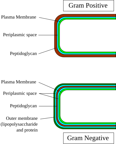
Gram staining takes advantage of this difference though the process below:
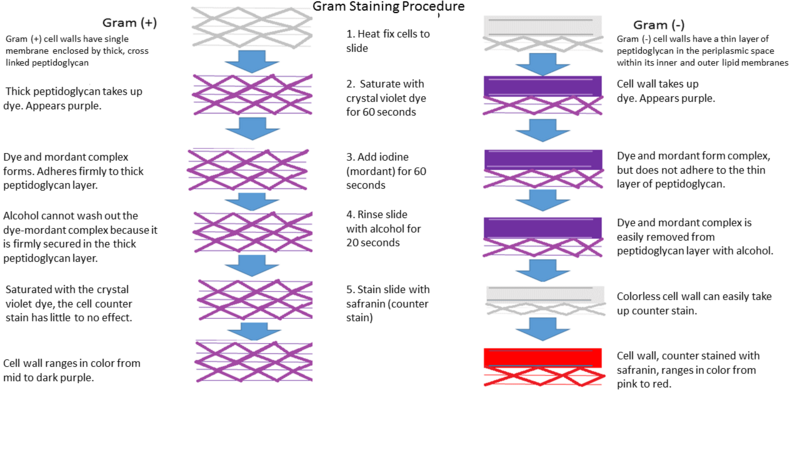
In the end, Gram-positive organisms will appear purple and Gram negative organisms will appear pink. Cells that are exposed too short or too long to the chemicals in the procedure can appear an intermediate color and cells that appear without a defined shape were likely damaged from heat exposure during the smear preparation.
Below is my first Gram stain. Is it Gram-positive or Gram-Negative?:

Correct! It is Gram-positive (the cell walls were purple under the microscope). Now it is time to examine the cell morphology. Lets take a look!

Unfortunately, this microscope did not have the ability to take pictures. But oh I wish I could share what I saw with you! This is the first time I have ever used so powerful a microscope and it was amazing. I sat there looking around, yes at the same identical clones, for almost 30 minutes (switching back and forth between 4x, 10x, and 40x magnification). While part of the time was getting used to using a microscope, the rest was marveling at this unseen world that exists all around us.
As for the project, I used this opportunity to describe the cell morphology of the unknown bacteria. Much like the colony morphology, different bacteria have characteristic patterns of appearance from which they can be identified.

The unknown bacteria was definitely a cocci and had bunching like staphylococci.
Before I left for the day I prepared one last test to verify the Gram-positive result. I prepared a MacConky plate. It looks just like the others except the medium is designed to inhibit the growth of Gram-positive organisms (using crystal violet and bile salts) which allows for the isolation of Gram-negative organisms which can multiply in such environments.

If the Gram-positive results is correct these should sit in the incubator and when I return they should look the same, with no growth.


Comments
Post a Comment